Tuesday, August 26, 2014

"Brooklyn’s Morbid Anatomy Museum: A New and Resplendent Temple of Weirdness," Ariella Budick, Financial Times, August 2014

One of us, one of us, we accept her, one of us, gooble gobble, one of us.” If you, too, are one of us – that is, a slightly embarrassed devotee of all things creepy and surreal – you might recognise that chant. It comes from Tod Browning’s 1932 horror film Freaks, as a troupe of circus sideshow performers welcomes a horrified new member.
A similar fearful camaraderie unites those who make their way to the Morbid Anatomy Museum, a new and resplendent temple of weirdness in New York. With its collection of oddities – a wax model of a man disfigured by syphilis, a mummified rodent, and so on – the museum speaks to a cluster of fascinations that has only tangentially to do with death.
Rather, its aficionados – and I speak from the heart, here – are attuned to the slightly bizarre aspects of ordinary existence. We respond to the nostalgic frisson emanating from surrealist photographs, 17th-century cabinets of curiosity, graphic histories of hysteria, faked photos of seances, studies of conjoined twins, and Coney Island in its tawdry heyday. We claim a distinguished roster of elders: Edgar Allan Poe, Edward Gorey, Georges Bataille, WG Sebald and Susan Sontag. I imagine this conclave of eccentrics gathered around a Ouija board, with Freud acting as master of ceremonies. His concept of the uncanny – what he called a “ghastly harbinger of death” – hovers invisibly throughout the museum.
This indispensable new institution was founded and funded by a cadre of likeminded obsessives, led by creative director Joanna Ebenstein.... offers classes in areas where art and morbidity overlap: how to articulate a snake skeleton; how to stuff, mount and costume rabbits in anthropomorphic poses; how to draw a human skull. Almost nightly lectures merge the outlandish with the scholarly. “Lizard Mummies and Giant Squid Tentacles” gets behind the scenes at the American Museum of Natural History, and “Industrial Ladies” examines early 19th-century department store wax mannequins. All this erudition supports the museum’s overarching point: that a concern with the once-alive and the eerily lifelike has long suffused modern culture.
--"Brooklyn’s Morbid Anatomy Museum: A new and resplendent temple of weirdness has opened in New York," Ariella Budick, Financial Times, August 2014
The Morbid Anatomy Museum has been very lucky to have received quite a bit of press attention since opening our doors almost two months ago, but none has so aptly and elegantly summed up the larger Morbid Anatomy ethos, aesthetic and community as Ariella Budick's wonderfully insightful piece in last weekend's Financial Times.

I have quoted the piece at length above; you can read it in its entirety by clicking here (note: you must sign up to read it, but its free and worth it!)

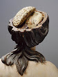
The photographs above are all courtesy of Stanley B Burns and the very excellent Burns Archive, one of the main loaners to our Art of Mourning exhibition, on view through December 4th.

If you would like to support The Morbid Anatomy Museum--wonderfully described in the article as an "indispensable new institution" (!)--please consider becoming a member (with all the benefits that entails!) here, or making a donation by clicking here

Monday, August 25, 2014

"Vanitas; the Transience of Earthly Pleasure" : Exhibition Catalog Review by Brenna Pladsen, Morbid Anatomy Docent and Volunteer

Vanitas: The Transience of Earthly Pleasures is an exhibition catalog for a Frieze Art Fair exhibition of the same name staged in 2010 by London's All Visual Arts Gallery. Morbid Anatomy Museum docent--and volunteer extraordinaire--Brenna Pladsen took a liking to the book, and kindly offered to write the following guest post about it; if you are interested in finding out more, you can spend some quality time with it at The Morbid Anatomy Library!
Vanitas is a prime example of a good exhibition book. This is partially because it seems to have been an excellent exhibition. Judging from the book, the art skewed toward the luxurious and the animal. Bits of birds and bugs are a striking part of the collection and bring home the concept of the vanitas painting, which is given a robust and generous explanation in the introduction.

The setting and curation are carefully matched to inspire the visitor into the correct frame of mind to appreciation the philosophy of the genre. The book follows that idea, and the wonderful photographs of the space can really transport you there and give a sense to the space and scale of the works. The Tim Noble and Sue Webster pieces, especially Metal Rats Fucking, need the accompanying context to be striking. The art is beautifully recorded by the photographs, but the exhibition photos are really transporting and have the dark, hyper-real quality of an original vanities painting. While the photography is luscious and luxurious, it is thankfully given space to breathe. I was struck by how contemporary and modern the book feels without loosing that decedent, velvety touch.

Tuesday, August 5, 2014

Plague Buboes and Preserved Primates – The Morbid Anatomy/Museum Vrolik “Amsterdam Weekend of Anatomy”: Guest Post by Schemenkabinett

The following is a review of the recent Morbid Anatomy/Museum Vrolik Amsterdam Weekend of Anatomy originally published by Katharina von Oheimb and Parm von Oheimb on their German-language Schemenkabinett blog. A translation of the piece, especially for Morbid Anatomy readers, follows; you can see the original piece by clicking here. To get on the Morbid Anatomy mailing list and thus be alerted to similar events in the future, click here.
In Amsterdam, we devoted ourselves to the field between anatomy and arts for a whole weekend.

He was called the “Artist of Death.” On the screen in the small lecture room at the Academic Medical Center in Amsterdam, a surreal scene appears, showing human fetal skeletons that seem to wipe away their tears with handkerchiefs made of preserved meninges. It is one of Frederick Ruysch’s (1638-1731) anatomical dioramas (top image). With his preparations and dioramas, Dutch anatomist and botanist Ruysch created unique works of art. The attendees in the lecture room look with fascination at his works. More than sixty participants have gathered at the weekend of 10th and 11th May 2014 to take part at the “Amsterdam Weekend of Anatomy” at Museum Vrolik. The event was brought into being by Joanna Ebenstein from Morbid Anatomy and Laurens de Rooy, the director of Museum Vrolik. Together with the other attendees--who came mostly came from the Netherlands, but also from Belgium, Germany, Great Britain, Poland, and the USA--we are listening to the first talks of this day. We will encounter Frederick Ruysch several times again during the next days, because his connection between scientific accuracy and artistic presentation can be discovered in every corner of this place.

Marieke Hendriksen reports in her talk about the puzzle of the beaded babies, which she investigated for her dissertation. An image on the screen shows a newborn baby in a jar filled with preservational liquid, decorated with beaded strings around its neck and wrists. In total, only eleven of such decorated specimens are known. All of them are from Dutch collections and from between 1780 and 1810. Hendriksen explains how she thoroughly investigated historical collections and how she examined literature to find evidence for the origin of these unique specimens. Probably, they stem from Dutch colonies; this puzzle, however, has not been finally solved yet.

In the afternoon, we participate in a wax modeling course (second image down). Medical wax models, so called moulages, are lifelike moldings of diseased body parts. They served for the training of physicians, but have also been used for explaining disease symptoms to the public. Due to the plasticity of the presentations, they have been clearly superior to drawings. For the creation of moulages, modeling material has often been applied directly to diseased parts of skin to obtain highly realistic moldings. In this way, whole series could be created, which documented the development of diseases or the effect of therapies. Only with the emergence of color photography, moulages became less important.
As we are entering the course room, pale wax faces already wait at our workplaces. During the next hours, we add disease symptoms and several wounds to them under the tutelage of Eleanor Crook. The London-based artist Crook has specialized in the creation of moulages. In courses like this, she explains impressively how plastic plague buboes can be formed from a special wax and later get colorized in bluish black. After finishing the course, we proudly bag our grotesque faces that are terribly disfigured by plague and syphilis and meet for the end of the evening at the exhibition room of Museum Vrolik, which has been exclusively opened for us. In the midst of all the showcases full of medical specimens, we have the opportunity to acquaint with the highly interesting group of participants.

The next day starts with a talk about the history of the moulages held by the Academic Medical Center in Amsterdam (third image down). These artfully designed moldings stem from the early 20th century. Soon afterwards, we find ourselves in the part of the building complex, where the moulages are stored today. Faces that are badly marked by diseases but appear strangely alive, as well as arms, hands, and genitals are stored in showcases and cabinets. Most of these medical artworks have been created at the island Sumatra and present tropical skin diseases.

Afterwards, the preparator Inge Dijkman gives us an introduction to her work at Museum Vrolik (bottom image). She has brought a small selection of specimens with her. Very cautious, Dijkman lifts an almost 10 cm large, preserved fetus out of the liquid and shows it to us. She explains that this fetus had implanted outside the uterine cavity. During such a so-called ectopic pregnancy, the fetus dies in most cases.
After this exceptional insight into the preparation laboratory, we take part at a guided tour to selected specimens of Museum Vrolik, where we learn about several fascinating details. Willem Vrolik (1801-1863), for example, one of the founders of the collection, was the first person that recorded multiple congenital disorders. He described, for instance, the first known case of the Smith-Lemli-Opitz syndrome. Individuals who suffer from this syndrome are not able to produce cholesterol. The external characteristics of the syndrome are diverse and range from female genitalia in combination with a male chromosome set, to malpositions of hands and feet, and to supernumerary fingers and toes. Depending on the severity of symptoms, where internal organs are also mostly limited in their functionality, this genetic disease often ends fatally.

At the end of our weekend at Museum Vrolik, a guided tour leads to the collection rooms in the basement of the Academic Medical Center, which are usually closed to the public. During the Cold War, a nuclear bombproof underground hospital was installed here. Today, the thick walls and heavy door locks bring the former function still to mind. Curator Laurens de Rooy guides us to the first room, which contains parts of the anthropological and zoological collections. In the second room, multiple specimen jars are stored in shelves. Animals with malformations are a main focus of the collection and have been used for comparative anatomy studies. In the third room, dry preparations are stored. Various preserved skulls of humans and other primates stand close together. One of our highlights in this room is the artfully created historical dry preparation of a human arm, where the pattern of tendons is clearly visible. By leaving the underground rooms, our “Amsterdam Weekend of Anatomy” ends.
During the two days at Museum Vrolik, we could gather various exciting impressions and meet a lot of interesting people. The event connected arts and science in a unique way and was worthwhile in all respects. Due to the high approval of the “Amsterdam Weekend of Anatomy,” the event is planned to take place again next year in a similar form.
Figures:
  1. Anatomical diorama by Frederik Ruysch (engraving by Cornelius Huyberts)
  2. In the course it is learned how medical max models are created. Photo courtesy of Katharina von Oheimb and Parm von Oheimb.
  3. Collection of historical moulages at the Academic Medical Center. Photo courtesy of Katharina von Oheimb and Parm von Oheimb.
  4. Insights into different preparation techniques. Photo courtesy of Katharina von Oheimb and Parm von Oheimb.